|
heidelberg engineering
ss-oct  Ss Oct, supplied by heidelberg engineering, used in various techniques. Bioz Stars score: 90/100, based on 1 PubMed citations. ZERO BIAS - scores, article reviews, protocol conditions and more https://www.bioz.com/product/ss-oct+system/ss+oct/pmc08770693-2-4-1 Average 90 stars, based on 1 article reviews
ss-oct - by Bioz Stars,
2026-09
90/100 stars
|
Buy from Supplier |
|
OptoRes GmbH
mhz-oct omes 4d mhz-oct system Mhz Oct Omes 4d Mhz Oct System, supplied by OptoRes GmbH, used in various techniques. Bioz Stars score: 90/100, based on 1 PubMed citations. ZERO BIAS - scores, article reviews, protocol conditions and more https://www.bioz.com/product/ss-oct+system/mhz+ss+oct+system/pmc08548019-280-8-2 Average 90 stars, based on 1 article reviews
mhz-oct omes 4d mhz-oct system - by Bioz Stars,
2026-09
90/100 stars
|
Buy from Supplier |
|
heidelberg engineering
ss-oct anterion  Ss Oct Anterion, supplied by heidelberg engineering, used in various techniques. Bioz Stars score: 90/100, based on 1 PubMed citations. ZERO BIAS - scores, article reviews, protocol conditions and more https://www.bioz.com/product/ss-oct+system/ss+oct+anterion/pmc07775111-15-4-7 Average 90 stars, based on 1 article reviews
ss-oct anterion - by Bioz Stars,
2026-09
90/100 stars
|
Buy from Supplier |
|
Movu Inc
swept-source optical coherence tomography biometer argos Swept Source Optical Coherence Tomography Biometer Argos, supplied by Movu Inc, used in various techniques. Bioz Stars score: 90/100, based on 1 PubMed citations. ZERO BIAS - scores, article reviews, protocol conditions and more https://www.bioz.com/product/ss-oct+system/argos+ss+oct+biometer/pmc11900115-24-10-19 Average 90 stars, based on 1 article reviews
swept-source optical coherence tomography biometer argos - by Bioz Stars,
2026-09
90/100 stars
|
Buy from Supplier |
|
Movu Inc
ss-oct-based biometry Ss Oct Based Biometry, supplied by Movu Inc, used in various techniques. Bioz Stars score: 90/100, based on 1 PubMed citations. ZERO BIAS - scores, article reviews, protocol conditions and more https://www.bioz.com/product/ss-oct+system/ss+oct+based+biometry/pm37525287-94-31-33 Average 90 stars, based on 1 article reviews
ss-oct-based biometry - by Bioz Stars,
2026-09
90/100 stars
|
Buy from Supplier |
|
topcon corporation
deep range imaging (dri) dri-triton ss-oct  Deep Range Imaging (Dri) Dri Triton Ss Oct, supplied by topcon corporation, used in various techniques. Bioz Stars score: 90/100, based on 1 PubMed citations. ZERO BIAS - scores, article reviews, protocol conditions and more https://www.bioz.com/product/ss-oct+system/deep+range+imaging++dri++triton+ss+oct+device/pmc07235989-73-15-21 Average 90 stars, based on 1 article reviews
deep range imaging (dri) dri-triton ss-oct - by Bioz Stars,
2026-09
90/100 stars
|
Buy from Supplier |
|
topcon corporation
oct-a Oct A, supplied by topcon corporation, used in various techniques. Bioz Stars score: 90/100, based on 1 PubMed citations. ZERO BIAS - scores, article reviews, protocol conditions and more https://www.bioz.com/product/ss-oct+system/ss+oct/pm29924197-64-9-15 Average 90 stars, based on 1 article reviews
oct-a - by Bioz Stars,
2026-09
90/100 stars
|
Buy from Supplier |
|
topcon corporation
dri oct triton device Dri Oct Triton Device, supplied by topcon corporation, used in various techniques. Bioz Stars score: 90/100, based on 1 PubMed citations. ZERO BIAS - scores, article reviews, protocol conditions and more https://www.bioz.com/product/ss-oct+system/ss+oct+device+dri+oct+triton/10__3390_slash_app13021152-99-10-14 Average 90 stars, based on 1 article reviews
dri oct triton device - by Bioz Stars,
2026-09
90/100 stars
|
Buy from Supplier |
|
topcon corporation
ss-oct topcon dti trition Ss Oct Topcon Dti Trition, supplied by topcon corporation, used in various techniques. Bioz Stars score: 90/100, based on 1 PubMed citations. ZERO BIAS - scores, article reviews, protocol conditions and more https://www.bioz.com/product/ss-oct+system/ss+oct+topcon+dti+trition/pm37498843-95-3-7 Average 90 stars, based on 1 article reviews
ss-oct topcon dti trition - by Bioz Stars,
2026-09
90/100 stars
|
Buy from Supplier |
|
EyeSys Laboratories
ss oct system Ss Oct System, supplied by EyeSys Laboratories, used in various techniques. Bioz Stars score: 90/100, based on 1 PubMed citations. ZERO BIAS - scores, article reviews, protocol conditions and more https://www.bioz.com/product/ss-oct+system/ss+oct+system/10__1364_slash_boe__2__002709-366-6-18 Average 90 stars, based on 1 article reviews
ss oct system - by Bioz Stars,
2026-09
90/100 stars
|
Buy from Supplier |
|
alcon inc
swept source optical coherence tomography biometer argos  Swept Source Optical Coherence Tomography Biometer Argos, supplied by alcon inc, used in various techniques. Bioz Stars score: 90/100, based on 1 PubMed citations. ZERO BIAS - scores, article reviews, protocol conditions and more https://www.bioz.com/product/ss-oct+system/argos+ss+oct+optical+biometer/pmc11594376-68-15-23 Average 90 stars, based on 1 article reviews
swept source optical coherence tomography biometer argos - by Bioz Stars,
2026-09
90/100 stars
|
Buy from Supplier |
|
Canon inc
ss-oct/octa device  Ss Oct/Octa Device, supplied by Canon inc, used in various techniques. Bioz Stars score: 90/100, based on 1 PubMed citations. ZERO BIAS - scores, article reviews, protocol conditions and more https://www.bioz.com/product/ss-oct+system/ss+oct+octa+device/bio_rxiv__2023__06__12__544445-169-18-22 Average 90 stars, based on 1 article reviews
ss-oct/octa device - by Bioz Stars,
2026-09
90/100 stars
|
Buy from Supplier |
Image Search Results
Journal: Scientific Reports
Article Title: High speed, long range, deep penetration swept source OCT for structural and angiographic imaging of the anterior eye
doi: 10.1038/s41598-022-04784-0
Figure Lengend Snippet: SS-OCT imaging performance. ( a ) Time-integrated spectrum of the MEMS-VCSEL light source. The booster optical amplifier driving current is modulated to achieve a symmetrical bell-shaped spectrum for improved sensitivity and sidelobe suppression. Arrows: spikes in spectral density occur because of the slowdown and reversal of the wavelength sweep. ( b ) The MEMS-VCSEL wavelength sweep with respect to time. The solid red line indicates difference between the measured sweep wavenumber (black) and ideal linear sweep (red dashed line). Shaded area represents the wavelength band used in OCT signal reconstruction. ( c ) OCT sensitivity versus imaging depth. Note that a constant sensitivity is maintained within the first ~ 13.5 mm axial range. Dashed lines correspond to a 6 dB drop in sensitivity. ( d ) OCT point spread functions from 4 imaging depths. The axial resolution is 12.2 µm full-width at half-maximum (FWHM) in air, and is invariant for imaging depths < ~ 13.5 mm.
Article Snippet: Anterion (
Techniques: Imaging

Journal: Scientific Reports
Article Title: High speed, long range, deep penetration swept source OCT for structural and angiographic imaging of the anterior eye
doi: 10.1038/s41598-022-04784-0
Figure Lengend Snippet: Wide field, long range, deep penetration structural imaging using the SS-OCT prototype instrument. ( a ) En face amplitude projection of a 25-mm OCT volume. ( b ) 6-mm horizontal B-scan centered at the limbus. Yellow arrow: trabecular meshwork. Red arrowhead: Schlemm’s canal. The connected hypo-reflective curvilinear feature may correspond to a collector vessel segment. ( c ) 6-mm horizontal B-scan 6 mm temporal to the corneoscleral junction. Blue arrowhead: insertion tendon of the lateral rectus muscle.
Article Snippet: Anterion (
Techniques: Imaging

Journal: Scientific Reports
Article Title: High speed, long range, deep penetration swept source OCT for structural and angiographic imaging of the anterior eye
doi: 10.1038/s41598-022-04784-0
Figure Lengend Snippet: SS-OCT imaging of the full anterior segment of the eye. ( a )–( c ) Extracted 20-mm B-scans where the focal depth position is dynamically adjusted with 18 steps at ~ 500 µm spacing. The focal plane shifts from the cornea to the posterior surface of the lens capsule. White arrow: corresponding location of the focal plane. ( d ) Fused full depth high resolution cross-sectional image using all focally stepped B-scans. ( e ) 6-mm OCT volume of an eye with subcapsular cataract. The focal plane is fixed at the posterior lens capsule. The B-scan corresponds to the horizontal meridian (white arrowhead). Insert: en face OCT amplitude projection along the depth range of the posterior half of the lens capsule. The lateral extent of the cataract lesion can be appreciated. Saccadic motion was manually corrected using rigid strip based registration.
Article Snippet: Anterion (
Techniques: Imaging, Stripping Membranes

Journal: Scientific Reports
Article Title: High speed, long range, deep penetration swept source OCT for structural and angiographic imaging of the anterior eye
doi: 10.1038/s41598-022-04784-0
Figure Lengend Snippet: OCTA imaging using the SS-OCT prototype instrument. ( a ) Digital image of the subject. Boxes indicates locations of the OCTA scans. ( b ) 9-mm OCTA projection, scanned at the location corresponding to the dashed box. ( c )–( e ) 6-mm OCTA projections, scanned at locations corresponding to the solid boxes, respectively. ( f )–( g ) OCTA B-scans, extracted respectively from the center of the imaging field. Note the highly scattering sclera generates strong vertical projection artifacts. The OCTA signal posterior to the sclera is assumed to originate from the choroid.
Article Snippet: Anterion (
Techniques: Imaging

Journal: Scientific Reports
Article Title: High speed, long range, deep penetration swept source OCT for structural and angiographic imaging of the anterior eye
doi: 10.1038/s41598-022-04784-0
Figure Lengend Snippet: Iris OCTA using the SS-OCT prototype instrument. ( a ) 9-mm and ( b ) 6-mm OCTA of an Asian eye (dark pigmented). ( c )–( d ) 12-mm OCTA of a Caucasian eye (lightly pigmented). ( c ) shows a dilated pupil in dark environment, where ( d ) shows a constricted pupil in response to white light stimulation. Iris vessels show morphological changes following pupillary constriction, where the vessels appear straighter and brighter. ( e , g ) Structural and ( f , h ) angiographic B-scans extracted from the corresponding scans at the locations indicated by the yellow arrowhead.
Article Snippet: Anterion (
Techniques:

Journal: Scientific Reports
Article Title: High speed, long range, deep penetration swept source OCT for structural and angiographic imaging of the anterior eye
doi: 10.1038/s41598-022-04784-0
Figure Lengend Snippet: Comparison of selected commercial OCT instruments and the SS-OCT prototype instrument. Axial resolution and imaging depth reported correspond to values in tissue.TD-OCT = time domain OCT.
Article Snippet: Anterion (
Techniques: Imaging

Journal: Scientific Reports
Article Title: High speed, long range, deep penetration swept source OCT for structural and angiographic imaging of the anterior eye
doi: 10.1038/s41598-022-04784-0
Figure Lengend Snippet: SS-OCT prototype instrument design. ( a ) Schematic of the high speed SS-OCT interferometer. BPD: balanced photo detector; DC: dispersion compensation glass plates; GS: galvanometer scanner; ISO: optical isolator; MZI: Mach–Zehnder interferometer; NC: not connected; PC: polarization controller; PIU: patient interface unit; SL: scan lens. ( b ) Simplified diagram showing dynamic focal plane adjustment using an electrically focus tunable lens. CL: collimating lens; ETL: electrically tunable lens; FP: focal plane. ( c ) Photo of the OCT patient interface unit with cover removed.
Article Snippet: Anterion (
Techniques:
Journal: PLoS ONE
Article Title: Comparison study of the axial length measured using the new swept-source optical coherence tomography ANTERION and the partial coherence interferometry IOL Master
doi: 10.1371/journal.pone.0244590
Figure Lengend Snippet: Mean value, mean difference and 95% limit of agreement of axial length in 175 eyes and anterior chamber depth in 104 eyes readings of the two biometry devices.
Article Snippet: We compared the two
Techniques:

Journal: PLoS ONE
Article Title: Comparison study of the axial length measured using the new swept-source optical coherence tomography ANTERION and the partial coherence interferometry IOL Master
doi: 10.1371/journal.pone.0244590
Figure Lengend Snippet: Mean value, mean difference, and 95% limit of agreement (LoA) of keratometric values (K1, K2, J0 and J45) between two biometry.
Article Snippet: We compared the two
Techniques:
Journal: Diagnostics
Article Title: Choroidal Changes of Long-Term Type 1 Diabetic Patients without Retinopathy
doi: 10.3390/diagnostics10040235
Figure Lengend Snippet: Agreement between subfoveal choroidal thickness (SFCT) measured with both devices in both groups. Control group (blue) showed a distribution following the equation for optical coherence tomography (SS-OCT) (μm) = 0.7752 × SS-OCT(μm) + 40.511, being R 2 = 0.6595 and DM1 group (red) SS-OCT (μm) = 0.6497 × SS-OCT(μm) + 75.542, being R 2 = 0.6462.
Article Snippet: Each individual was imaged using a Spectralis SD-OCT (Heidelberg Engineering, Inc., Heidelberg, Germany) device and
Techniques: Tomography

Journal: Diagnostics
Article Title: Choroidal Changes of Long-Term Type 1 Diabetic Patients without Retinopathy
doi: 10.3390/diagnostics10040235
Figure Lengend Snippet: Choroidal thicknesses in the ETDRS quadrants expressed in μm (from the Bruch’s Membrane (BM) to the choroid-scleral limit) measured with the Spectralis spectral domain optical coherence tomography (SD-OCT) and the deep range imaging (DRI)-Triton swept source optical coherence tomography (SS-OCT) in the type 1 diabetes Mellitus (DM1) group and the control group. Values are presented as the mean ± standard deviation and range (minimum-maximum). Significant differences were considered as p < 0.05, calculated by Kolmogorov–Smirnov test for two independent nonparametric samples, are shown with *. The volume is represented in mm 3 .
Article Snippet: Each individual was imaged using a Spectralis SD-OCT (Heidelberg Engineering, Inc., Heidelberg, Germany) device and
Techniques: Tomography, Imaging, Standard Deviation

Journal: Diagnostics
Article Title: Choroidal Changes of Long-Term Type 1 Diabetic Patients without Retinopathy
doi: 10.3390/diagnostics10040235
Figure Lengend Snippet: Mean of choroidal thicknesses in each ETDRS area expressed in μm measured with the Spectralis spectral domain optical coherence tomography (SD-OCT) and the deep range imaging (DRI)-Triton swept source optical coherence tomography (SS-OCT) in the type 1 diabetes Mellitus (DM1) group and the control group. Significant differences were considered as p < 0.05, calculated by Kolmogorov–Smirnov test for two independent nonparametric samples, are shown with *. SD-OCT, spectral domain optical coherence tomography; DRI, deep range image; SS-OCT, swept source optical coherence tomography; DM, diabetes mellitus; ETDRS, Early Treatment Diabetic Retinopathy Study; R1, central; S, superior; T, temporal; I, inferior; N, nasal. The number 1 corresponds to the 3 mm parafoveal inner circle and 2 to the 6 mm perifoveal outer circle.
Article Snippet: Each individual was imaged using a Spectralis SD-OCT (Heidelberg Engineering, Inc., Heidelberg, Germany) device and
Techniques: Tomography, Imaging

Journal: Diagnostics
Article Title: Choroidal Changes of Long-Term Type 1 Diabetic Patients without Retinopathy
doi: 10.3390/diagnostics10040235
Figure Lengend Snippet: Choroidal thicknesses by ETDRS quadrants in μm (from the BM to the choroid-scleral limit) assessed with Spectralis spectral domain optical coherence tomography (SD-OCT) and the deep range imaging (DRI)-Triton swept source optical coherence tomography (SS-OCT) in the type 1 diabetes Mellitus (DM1) group subdivided into two groups: less than 24 years and over or equal to 24 years of evolution of DM1. Values are presented as the mean ± standard deviation and range (minimum–maximum). Significant differences were considered as p < 0.05, calculated by Kolmogorov–Smirnov test for two independent nonparametric samples, are shown with *. The volume is represented in mm 3 .
Article Snippet: Each individual was imaged using a Spectralis SD-OCT (Heidelberg Engineering, Inc., Heidelberg, Germany) device and
Techniques: Tomography, Imaging, Standard Deviation

Journal: Diagnostics
Article Title: Choroidal Changes of Long-Term Type 1 Diabetic Patients without Retinopathy
doi: 10.3390/diagnostics10040235
Figure Lengend Snippet: Mean of choroidal thicknesses in each ETDRS area in μm (from the BM to the choroid-scleral limit) assessed with Spectralis spectral domain optical coherence tomography (SD-OCT) and the deep range imaging (DRI)-Triton swept source optical coherence tomography (SS-OCT) in the type 1 diabetes Mellitus (DM1) group subdivided into two groups: less than 24 years and over or equal to 24 years of evolution of DM1. Significant differences were considered as p < 0.05, calculated by Kolmogorov–Smirnov test for two independent nonparametric samples, are shown with *. SD-OCT, spectral domain optical coherence tomography; DRI, deep range image; SS-OCT, swept source optical coherence tomography; DM, diabetes mellitus; ETDRS, Early Treatment Diabetic Retinopathy Study; R1, central; S, superior; T, temporal; I, inferior; N, nasal. The number 1 corresponds to the 3 mm parafoveal inner circle and 2 to the 6 mm perifoveal outer circle.
Article Snippet: Each individual was imaged using a Spectralis SD-OCT (Heidelberg Engineering, Inc., Heidelberg, Germany) device and
Techniques: Tomography, Imaging
Journal: Journal of Clinical Medicine
Article Title: Efficacy of Sutureless Scleral Fixation of One-Piece T-Shaped Haptic Intraocular Lens in Maintaining Anterior Chamber Stability During Descemet Membrane Endothelial Keratoplasty in Vitrectomized Eyes: Leak Test and Iris Diaphragm Reconstruction
doi: 10.3390/jcm13226654
Figure Lengend Snippet: Case#2 of Descemet membrane endothelial keratoplasty (DMEK). ( A ) Slit-lamp color photograph at baseline and ( B ) at 3 months after DMEK surgery. ( C ) Reconstruction combining ultrasound biomicroscopy and anterior segment optical coherence tomography revealing both lens position and the attached DMEK graft 3 months after surgery.
Article Snippet: Preoperative measurements of axial length (AXL) and AC depth (ACD) were performed using a swept
Techniques: Membrane, Tomography
Journal: bioRxiv
Article Title: The orientation of choroidal macrophage polarization significantly influences the development of myopia in murine models
doi: 10.1101/2023.06.12.544445
Figure Lengend Snippet: (A) Three-weeks-old wild-type C57BL/6JJc1 mice were divided into two groups (4 per group unless otherwise indicated) and one group was intraperitoneally injected with 10 ul/g LPS and the other with PBS. The injection lasted for 2 weeks, and refraction, axial length, and choroidal thickness were measured weekly with an infrared photorefractor and SD-OCT system. Choroidal blood perfusion was measured with OCTA. In addition, choroid samples were collected for real-time PCR. (B) Compared to the PBS injection group, the LPS injection group showed a significantly larger refractive change ( P < 0.001), significant axial eye growth ( P < 0,001), and substantially thinner choroid ( P < 0.001). Bars represent mean +/− standard deviations. (C) The choroidal blood perfusion decreased significantly after 2 weeks of LPS injection, compared to the PBS injection group. (D) mRNA levels of Tnf-α and IL-6 significantly increased after 2 weeks of LPS injection. P values were obtained from a t-test. * P < 0.05. ** P < 0.01. *** P < 0.001.
Article Snippet: Choroidal blood perfusion was measured at the beginning (3-week-old) and end (6-week-old) stages of myopia induction using an
Techniques: Injection, Real-time Polymerase Chain Reaction
Journal: bioRxiv
Article Title: The orientation of choroidal macrophage polarization significantly influences the development of myopia in murine models
doi: 10.1101/2023.06.12.544445
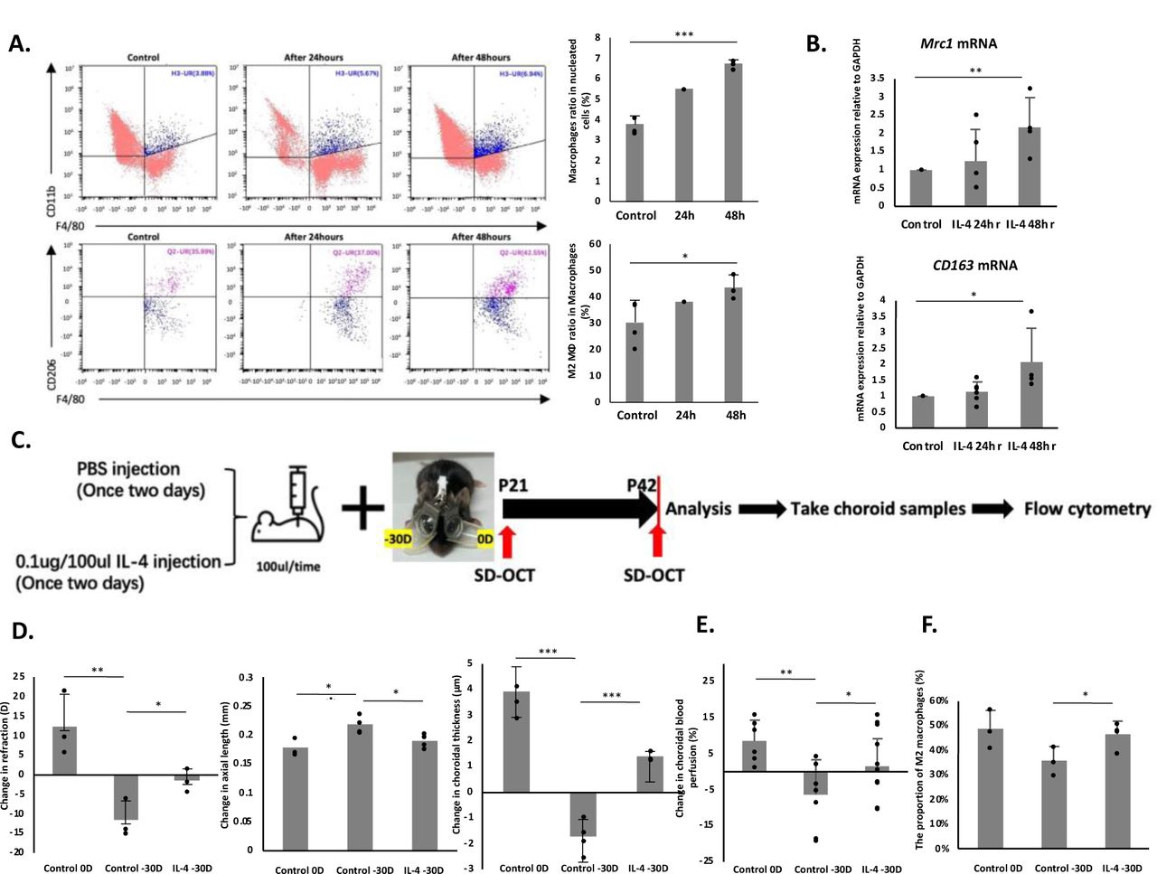
Figure Lengend Snippet: Three-week-old wild-type C57BL/6JJc1 mice were injected with 0.1 ug/100 ul IL-4, and choroid samples were collected at 0 h, 24 h, and 48 h after injection for flow cytometry and real-time PCR. (A) Macrophages in the H3-UR region were denoted as CD11b + and F4/80 + . The proportion of macrophages increased significantly after IL-4 injection ( P < 0.001). M2 macrophages in the Q2-UR and were identified as CD206 + . At 48 h after IL-4 injection, M2 macrophages were dramatically polarized ( P < 0.05). (B) Real-time PCR results showed that the mRNA expression levels of Mrc1 and CD163 increased significantly 48 h after IL-4 injection. (C) Three-weeks-old wild-type C57BL/6JJc1 mice were divided into two groups and injected with either 0.1 ug/100 ul IL-4 or PBS on alternate days with myopia induction. Refraction, axial length, and choroidal thickness were measured at the initial (3-week-old) and final (6-week-old) stages of myopic induction using an infrared photorefractor and SD-OCT system. (D) Eyes treated with −30D lenses showed a significantly larger refractive change ( P < 0.01), greater axial length elongation ( P < 0.05), and increased choroidal thickness ( P < 0.001) than those treated with 0D lenses in the PBS injection mice. Mice injected with 0.1 ug/100 ul IL-4 showed a significantly smaller refractive change ( P < 0.05), smaller change in axial length( P < 0.05), and positive change in choroidal thickness with a statistical significance ( P < 0.001). Bars represent the mean +/− standard deviation. (E) To evaluate changes in choroidal blood perfusion, three-weeks-old mice were divided into three groups: both eyes with 0D lenses (control 0D group), binocular myopia induction (control −30D group), and binocular myopia induction with IL-4 injection once every two days group (IL-4 −30D group). Choroidal blood perfusion was assessed at the initial (3-week-old) and end (6-week-old) stages of myopic induction using OCTA. Compared to the control 0D group, choroidal blood perfusion was significantly decreased in the control −30D group. Accordingly, compared with the control −30D group, choroidal blood perfusion was remarkably improved in the IL-4 −30D group. (F) Mice used to measure choroidal blood perfusion were euthanized at 6 weeks of age, and the choroids were collected for flow cytometry. The results showed that mice treated with −30D lenses and injected with IL-4 had a significant proportion of M2 macrophages compared to those treated with −30D lenses and injected with PBS. P values correspond to comparisons made using one-way ANOVA. * P < 0.05. ** P < 0.01. *** P < 0.001.
Article Snippet: Choroidal blood perfusion was measured at the beginning (3-week-old) and end (6-week-old) stages of myopia induction using an
Techniques: Injection, Flow Cytometry, Real-time Polymerase Chain Reaction, Expressing, Standard Deviation

Journal: bioRxiv
Article Title: The orientation of choroidal macrophage polarization significantly influences the development of myopia in murine models
doi: 10.1101/2023.06.12.544445
Figure Lengend Snippet: (A) Choroid samples were collected after 48 h of IL-13 injection and PBS injection for flow cytometry analysis, which was designed to evaluate the optimal time for the polarization of M2 macrophages. (B) Flow cytometry analysis demonstrated that both macrophage ratio and M2 macrophage ratio were significantly increased 48 h after IL-13 injection, compared to the PBS injection group. (C) Three-week-old mice were divided into three groups: 0D lenses on both eyes (control 0D group), binocular myopia induction (control −30D group), and binocular myopia induction and IL-13 injection (IL-13 −30D group). IL-13 injection frequency was once every two days. Refraction, axial length, and choroidal thickness were measured at the initial (3-week-old) and end (6-week-old) stages of myopic induction with an infrared photorefractor and SD-OCT system. (D) The control −30D group showed a significantly larger refractive change ( P < 0.001), greater axial length elongation ( P < 0.05), and thinner choroidal ( P < 0.001) compared to the control 0D group. The IL-13 −30D group indicated a significantly smaller refractive change ( P < 0.001), lesser axial length change ( P < 0.05), and positive change in choroidal thickness, with a statistical significance ( P < 0.001). Bars represent mean +/− standard deviations. (E) Choroidal blood perfusion was measured using OCTA. Compared to the control 0D group, choroidal blood perfusion was significantly decreased in the control −30D group. Correspondingly, compared to the control −30D group, choroidal blood perfusion was significantly improved in the IL-13 −30D group. (F) The proportion of M2 macrophages in the choroid showed a significant increase in the IL-13 −30D group, compared to the control −30D group. P values indicate comparisons relative to control with one-way ANOVA or t -test. * P < 0.05. ** P < 0.01. *** P < 0.001.
Article Snippet: Choroidal blood perfusion was measured at the beginning (3-week-old) and end (6-week-old) stages of myopia induction using an
Techniques: Injection, Flow Cytometry